Research Blog
IGF-1 LR3 vs IGF-1 DES: How Two Engineered IGF-1 Variants Differ in Research
Published
A structural comparison of IGF-1 LR3 and IGF-1 DES — how each engineered variant reduces IGF-binding-protein affinity and what published assays report.
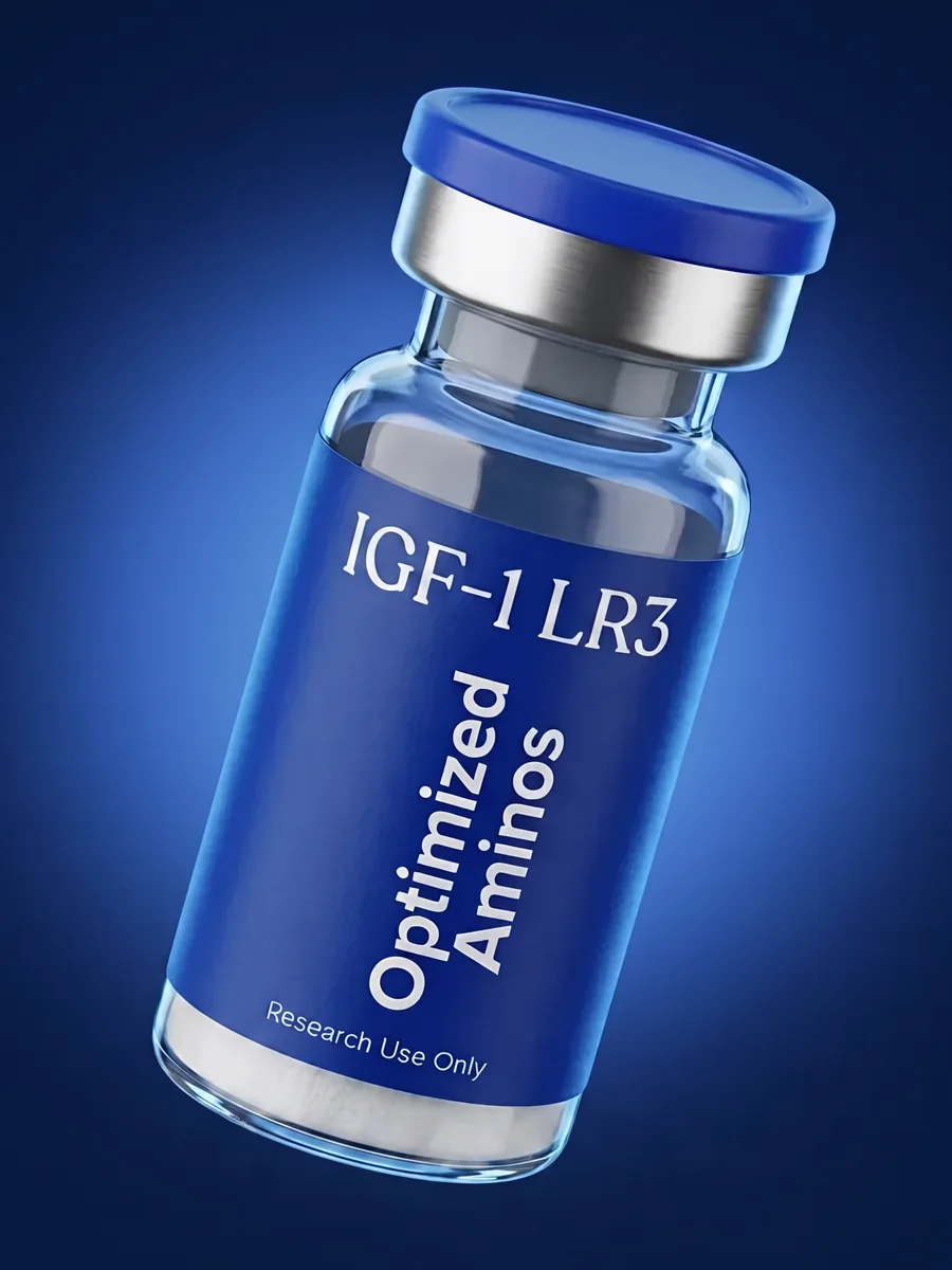
IGF-1 LR3 and IGF-1 DES are both laboratory-engineered variants of insulin-like growth factor 1 (IGF-1), designed by researchers to answer a specific methodological problem: native IGF-1 binds tightly to a family of IGF-binding proteins (IGFBPs) in serum and cell-culture media, which complicates in vitro experiments aimed at isolating IGF-1 receptor signaling itself. Both variants modify the native sequence to reduce IGFBP interaction — but they do it through different structural changes, and published research describes measurably different in vitro behavior as a result. This article compares the two variants side by side: their sequence differences, how each is characterized in binding and stability assays, and what published model-system data report about each.
Key Facts
- IGF-1 LR3 is native IGF-1 modified with a 13-amino-acid B-chain substitution plus an added arginine residue at position 3 (hence "Long R3").
- IGF-1 DES is native IGF-1 with the first three N-terminal amino acids (Gly-Pro-Glu) removed entirely — a truncation, not a substitution.
- Both variants are studied for reduced IGF-binding-protein (IGFBP) affinity relative to native IGF-1, but via different structural mechanisms.
- Published in vitro stability assays report a longer measured half-life for IGF-1 LR3 in cell-culture-medium conditions than for IGF-1 DES.
- Both variants retain measurable binding activity at the IGF-1 receptor (IGF-1R) in published receptor-binding assays, despite their structural modifications.
Why Engineered IGF-1 Variants Exist
Native IGF-1 is a 70-amino-acid single-chain polypeptide structurally related to proinsulin, with A, B, C, and D domains. In circulation and in most cell-culture systems, the majority of IGF-1 is bound to one of six known IGF-binding proteins (IGFBP-1 through IGFBP-6), which regulate its availability to the IGF-1 receptor. For researchers studying IGF-1 receptor signaling specifically — as opposed to the combined effect of IGF-1 plus its binding-protein regulation — this creates a confound: native IGF-1 added to a cell-culture assay is substantially sequestered by IGFBPs present in serum-supplemented media, making it difficult to isolate receptor-level signaling data. IGF-1 LR3 and IGF-1 DES were both developed, through different structural approaches, to reduce this IGFBP sequestration and give researchers a cleaner tool for IGF-1R pathway study.
IGF-1 LR3: The Substitution Approach
IGF-1 LR3 ("Long Arg3-IGF-1") modifies native IGF-1 in two ways: it substitutes a 13-amino-acid sequence from the human IGF-1 B-domain N-terminus, and it adds an extra arginine at position 3. Published binding-affinity studies report that this combined modification substantially reduces the molecule's affinity for IGFBPs compared to native IGF-1, while receptor-binding assays show it retains activity at the IGF-1 receptor. Our companion article, how IGF-1 LR3 was engineered for extended half-life, goes into more depth on the specific structural rationale behind the R3 substitution and what stability assays report about it.
Half-Life Characteristics in Published Assays
Because IGF-1 LR3 interacts less with IGFBPs, in vitro stability studies report it persists measurably longer in cell-culture-medium conditions than native IGF-1 or IGF-1 DES — a property researchers link to reduced IGFBP-mediated clearance and altered protease accessibility. For a broader explanation of how "half-life" is defined and measured in peptide research generally (rather than as a variant-specific claim), see our article peptide half-life explained for research contexts.
IGF-1 DES: The Truncation Approach
IGF-1 DES ("des(1-3)-IGF-1") takes a different structural route: rather than substituting or adding residues, it removes the first three N-terminal amino acids of native IGF-1 (glycine, proline, glutamic acid) entirely. This N-terminal tripeptide sits in a region of the molecule implicated in IGFBP interaction, so published binding studies report that its removal also reduces IGFBP affinity — but the resulting molecule is described in the literature as behaving somewhat differently from IGF-1 LR3 in comparative in vitro assays, including in reported binding-protein affinity magnitude and in cell-culture stability measurements.
Structural Comparison Summary
The core methodological distinction researchers cite between the two variants is substitution versus truncation:
- IGF-1 LR3 — B-domain 13-residue substitution + Arg addition at position 3; native sequence length is altered, not shortened.
- IGF-1 DES — N-terminal 3-residue deletion; the resulting peptide is shorter than native IGF-1 by three amino acids.
Both approaches converge on the same experimental goal — reduced IGFBP interference — but published comparative assays report they are not interchangeable in magnitude or in downstream stability characteristics, which is why researchers select one or the other based on the specific variable a given protocol is designed to isolate.
Receptor Binding: What's Retained
Despite their modifications, both IGF-1 LR3 and IGF-1 DES retain measurable binding activity at the IGF-1 receptor in published radioligand and cell-based competitive-binding assays, which is the property that makes them useful as IGF-1R-signaling research tools in the first place. Researchers designing IGF-1R pathway studies typically select between the two variants based on which structural trade-off — the LR3 substitution's extended in vitro persistence, or the DES truncation's shorter native-length backbone — best fits their specific assay design and controls.
Preparing Research Stock Solutions
As with any lyophilized peptide, preparing a research stock solution from either variant is a matter of concentration math: peptide mass (mg) divided by diluent volume (mL) yields the stock concentration in mg/mL. Because IGF-1 variants are typically supplied in small vial quantities (commonly 1 mg), precise measurement of diluent volume matters for downstream assay consistency. Our reconstitution calculator automates this calculation for a given vial size and diluent volume, which is useful when documenting stock-solution records for either variant.
Our IGF-1 LR3 research product page is the listing for researchers designing comparative or standalone IGF-1R pathway studies.
Frequently Asked Questions
What is the structural difference between IGF-1 LR3 and IGF-1 DES?
IGF-1 LR3 is native IGF-1 with a 13-amino-acid substitution at the B-chain N-terminus and an added Arg residue (the "long R3" modification), while IGF-1 DES has the first three N-terminal amino acids of native IGF-1 removed entirely, a truncation rather than a substitution.
Why do researchers study IGF-1 LR3's reduced binding-protein affinity?
IGF-1 LR3's structural modification is reported in binding-affinity studies to reduce its interaction with IGF-binding proteins (IGFBPs) compared to native IGF-1, which researchers use as a tool to isolate IGF-1 receptor signaling from IGFBP-mediated regulation in in vitro model systems.
Why is IGF-1 DES studied for reduced binding-protein interaction?
The N-terminal tripeptide removed in IGF-1 DES corresponds to a region implicated in IGFBP interaction, so published binding assays report that this truncation similarly reduces IGFBP affinity relative to native IGF-1, though through a different structural mechanism than the LR3 substitution.
Do IGF-1 LR3 and IGF-1 DES have different reported half-lives in model systems?
Published in vitro stability data describe IGF-1 LR3 as having a longer measured half-life in cell-culture-medium assays than IGF-1 DES, which researchers attribute to differences in how each variant's structural modification affects protease susceptibility and binding-protein interaction.
For laboratory and research use only. Not for human consumption.